
Hypodontia
What it means when teeth are missing from birth, how it is managed, and why early diagnosis changes everything
Not everyone develops a full set of teeth. For some people, one or several teeth simply never form: they are absent not because they have fallen out or been extracted, but because the developmental signal that should have triggered their formation either did not occur or failed partway through the process.
This is hypodontia, one of the most common dental developmental anomalies, and one that is frequently underdiagnosed until it presents as a clinical problem rather than being identified and managed from the outset.
Understanding hypodontia well enough to manage it effectively requires knowing which teeth are most commonly affected, what the clinical consequences are when they are absent, how retained primary teeth behave over time, and what the restoration options are once the diagnosis is established and the patient has finished growing.
At St James Dental Surgery in Muswell Hill, London, led by Dr Neha Tailor, whose background in biomimetic dentistry informs how missing and compromised teeth are approached restoratively, we assess and manage hypodontia across all age groups. Here is everything worth understanding about the condition.
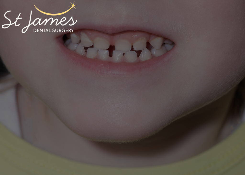
What hypodontia actually is: definitions and prevalence
Hypodontia is defined as the congenital absence of between one and five teeth, not including the third molars (wisdom teeth). Related terms cover the more severe ends of the spectrum:
Oligodontia is the congenital absence of six or more teeth, excluding third molars. This is a more severe presentation often associated with genetic syndromes and requiring more comprehensive multidisciplinary management.
Anodontia is the complete absence of all teeth, a very rare condition almost always associated with a specific genetic syndrome such as ectodermal dysplasia.
Hyperdontia is the opposite condition: the development of supernumerary (extra) teeth beyond the normal complement.
The prevalence of hypodontia varies between studies depending on the population examined and whether third molars are included. Excluding wisdom teeth, hypodontia affects approximately 2.5 to 6.9% of the general population. Including third molars, the figure rises to around 20 to 25%. This makes it one of the most common developmental dental anomalies, though many patients are unaware they have it, particularly where the absent teeth are those that would not have been visible anyway.
Which teeth are most commonly absent
Not all teeth are equally likely to fail to develop. The pattern of absence in hypodontia is consistent across populations, with certain teeth significantly more prone to agenesis (failure to develop) than others.
The most commonly absent teeth, in order of frequency:
- Third molars (wisdom teeth): By far the most frequently absent, affecting 20 to 25% of the population. The clinical significance varies considerably: absent wisdom teeth are often a clinical convenience rather than a management challenge.
- Mandibular second premolars (lower second premolars): The most commonly absent tooth in the non-wisdom-tooth category. They develop relatively late and appear to be more susceptible to the genetic disruptions that cause hypodontia.
- Maxillary lateral incisors (upper lateral incisors): The second upper front teeth (the smaller teeth either side of the central incisors). Absence here has significant aesthetic consequences, because these are visible teeth in the smile zone. The prevalence of congenital absence of upper lateral incisors is approximately 1.5 to 2% of the population, making it a condition that clinicians working in this area encounter regularly.
- Maxillary second premolars (upper second premolars): Less commonly absent than lower second premolars but within the same pattern.
- Mandibular central incisors (lower front teeth): Less common than the above, but clinically significant when present.
A key clinical feature: hypodontia is frequently bilateral and often symmetrical, affecting the same tooth on both sides of the arch. A patient with a missing upper right lateral incisor has a significantly elevated probability of a missing upper left lateral incisor as well.
The genetics and causes of hypodontia
Hypodontia has a strong genetic basis. Multiple genes involved in tooth development have been implicated, including PAX9, MSX1, AXIN2 and IRF6. Mutations in these genes affect the signalling pathways that control the initiation, morphogenesis and differentiation of tooth germs during early development.
The condition follows both autosomal dominant and autosomal recessive inheritance patterns depending on the specific genetic variant. This means that a patient with hypodontia has a meaningfully elevated probability of having affected family members, and their own children are at elevated risk compared to the general population.
Hypodontia can occur as an isolated developmental variation or as part of a broader syndrome:
- Ectodermal dysplasia: A group of conditions affecting the ectodermally derived tissues (skin, hair, nails, sweat glands and teeth). Oligodontia is a characteristic feature, often severe and affecting both primary and permanent dentitions.
- Down syndrome (trisomy 21): Associated with hypodontia in approximately 50% of cases, typically affecting the permanent dentition more than the primary dentition.
- Cleft lip and palate: The developmental disruption of the palatal and lip tissues is associated with hypodontia, particularly in the region adjacent to the cleft.
- Van der Woude syndrome: A condition involving lower lip pits, cleft lip and palate, and hypodontia.
Where hypodontia is identified in childhood, assessment for associated syndromic features is part of a thorough evaluation, particularly in cases of oligodontia or where multiple family members are affected.
Environmental factors have also been implicated: thalidomide exposure during the developmental period, chemotherapy or radiotherapy during tooth formation, and possibly low birth weight and maternal smoking are associated with increased hypodontia prevalence.
What happens when a tooth never develops: the clinical consequences
The absence of a tooth is not simply a gap. It creates a cascade of clinical consequences that develop progressively and become more significant over time if not managed.
Retained primary teeth
When a permanent tooth never develops, the primary (baby) tooth that would have been replaced often remains in the mouth for years or decades beyond its expected lifespan. A retained primary tooth is not a tooth substitute: its roots resorb unpredictably, its crown progressively wears, and the infrastructure that the permanent tooth would have laid down in terms of bone support is not created.
Retained primary teeth can provide functional and aesthetic service for many years, sometimes well into adulthood, but their long-term prognosis is uncertain and requires monitoring. Regular dental check-ups to assess the condition of retained primary teeth and the bone beneath them are an important part of hypodontia management.
Alveolar bone deficiency
The jawbone in the region of a missing tooth does not develop to its full volume without the stimulating presence of a tooth root. Where a tooth has never developed, the alveolar bone in that area is often deficient in both height and width. This has significant implications for any future implant placement, because implants require adequate bone volume for stability: a site with congenitally absent bone may require bone augmentation before an implant can be placed.
Mesial drift and space loss
The teeth adjacent to an edentulous space drift gradually toward it. Without management, the space that existed where the absent tooth should have been reduces, making future restoration more difficult or requiring orthodontic treatment to re-open the space before restoration.
Malocclusion
The absence of specific teeth, particularly posterior teeth involved in the bite, can produce malocclusions that affect both function and aesthetics. Missing upper lateral incisors allow the canines to drift forward, producing a specific appearance where the canine occupies the lateral incisor position, which is both aesthetically noticeable and functionally relevant.
Psychological impact
Particularly in younger patients, the absence of visible teeth, especially upper lateral incisors, has a meaningful aesthetic impact during the years when self-image is forming. This is not a trivial concern: it is a clinically relevant factor in the timing of treatment decisions.
The management of hypodontia: a multidisciplinary process
Hypodontia rarely has a single-step solution. Its management is almost always multidisciplinary, involving coordination between the general dentist, orthodontist, and restorative clinician, and its timing is governed by the patient’s stage of dental and skeletal development.
The role of early diagnosis
Identifying hypodontia as early as possible is clinically significant because it allows space management decisions to be made at the right time, before space has closed, before bone has resorbed further, and before primary teeth have failed. Routine radiographic assessment during children’s dental appointments is the mechanism by which absent permanent teeth are identified in childhood.
Panoramic X-rays (OPGs) taken at around the age of seven to eight, when the developing permanent teeth are visible radiographically, reveal which permanent teeth are and are not forming. This is the point at which the absence of permanent teeth should be identified and a management plan initiated.
At St James Dental Surgery, children’s dental appointments are not simply cavity checks. They include assessment of dental development and eruption, appropriate radiographic monitoring, and early identification of developmental anomalies including hypodontia, allowing referral and planning to begin at the optimal time.
Orthodontic space management
Orthodontic treatment plays two possible roles in hypodontia management, and the choice between them is one of the most significant decisions in the treatment plan.
Space closure: The spaces left by absent teeth are closed orthodontically, distributing the dentition so that the remaining teeth fill the arch without gaps. For some presentations of missing lower premolars, this produces a functional, aesthetic outcome without any prosthetic replacement.
Space opening: Where the remaining primary tooth has been or will be lost and a prosthetic replacement is planned, orthodontics opens and maintains the space to the correct dimensions for a restoration. This requires careful timing and coordination with the restorative phase.
Restorative options: what is available and when
The restorative management of hypodontia is determined by the patient’s age, the site of the absent tooth, the bone volume available, and the patient’s overall dental health.
Composite bonding: For missing upper lateral incisors where the adjacent teeth have been positioned correctly by orthodontics, reshaping the canines (which have moved into the lateral position) with composite bonding can produce a natural-looking result without any tooth replacement. This is sometimes called “canine substitution.” The canine is reshaped to look like a lateral incisor, and the first premolar is adjusted to look like the canine.
Dr Neha Tailor’s background in biomimetic dentistry is directly relevant here: biomimetic principles applied to composite bonding on canine substitution focus on replicating not just the appearance but the optical and mechanical properties of the tooth being mimicked, producing restorations that function naturally within the restored arch.
Dental bridges: Where space has been opened and bone levels are appropriate, a conventional bridge (supported by the adjacent teeth) or a resin-bonded bridge (bonded to the back of an adjacent tooth with a wing rather than requiring crown preparation) can replace a missing tooth permanently. Resin-bonded bridges are particularly appropriate for lateral incisor replacement in younger patients for whom implants are not yet indicated.
Dental implants: The gold standard for tooth replacement in most adult presentations of hypodontia. An implant placed into the bone at the site of the absent tooth replicates the root function, stimulates the bone, and supports a crown that is indistinguishable from the natural tooth it replaces. The critical timing consideration: implants should not be placed until jaw growth is complete, typically around the age of eighteen in females and nineteen to twenty in males. Placing an implant before growth is complete results in the implant being left behind as the jaw grows, producing an aesthetic complication that is difficult to correct.
Dentures and partial dentures: For extensive oligodontia or where implants are not feasible, partial or complete dentures provide removable replacement teeth. Modern denture designs are considerably more natural-looking than older versions, and for patients with significant tooth absence they remain a practical, accessible solution.
The role of ongoing maintenance in hypodontia
Whatever the restorative pathway chosen for hypodontia, ongoing maintenance is essential.
Retained primary teeth need regular monitoring to assess root resorption and crown condition. Dental hygienist appointments maintain the gum health around retained primary teeth and any prosthetic restorations, reducing the risk of gum disease at sites that may already have reduced bone support. Implants require specific cleaning around the implant margin: peri-implantitis, the implant equivalent of gum disease, is a risk that regular professional maintenance significantly reduces.
Regular dental check-up appointments allow the clinical team to monitor the condition of retained primary teeth, assess the stability of any restorations, and identify any emerging issues early. For hypodontia patients, these appointments carry particular clinical importance because the dental situation is inherently more complex than average and more susceptible to change over time.
The bottom line
Hypodontia is a common developmental condition affecting the number of teeth a person develops, most frequently involving lower second premolars, upper lateral incisors and third molars. Its management is multidisciplinary, time-sensitive and shaped by the specific clinical presentation: which teeth are absent, what the remaining dentition looks like, what bone is available, and where the patient is in their development.
Early identification, through routine children’s dental appointments with appropriate radiographic monitoring, allows space management decisions to be made at the right time and restorative planning to be initiated before the clinical situation becomes more complex.
At St James Dental Surgery in Muswell Hill, Dr Neha Tailor provides thorough assessment, biomimetically informed restorative planning, and ongoing management for patients with hypodontia at all stages. For appointments, call 020 8365 2090 or visit us at 18 Muswell Hill Broadway, London N10 3RT.
For urgent concerns about dental development or related symptoms, our emergency dental service in Muswell Hill is available for same-day assessment.
Disclaimer
The information in this article is intended for general educational guidance only and does not constitute personalised dental advice. For concerns about missing teeth or dental development, please book an appointment with a qualified dental professional for a proper clinical assessment.
St James Dental Surgery is a private dental practice at 18 Muswell Hill Broadway, London N10 3RT, led by Dr Neha Tailor. We offer dental check-ups, children’s dentistry, dental hygienist appointments, emergency dental care, biomimetic dentistry, dental implants, Invisalign, composite bonding, porcelain veneers, teeth whitening, dental crowns and smile makeovers. Call 020 8365 2090.
Frequently asked questions
How is hypodontia diagnosed?
Hypodontia is most reliably diagnosed through dental radiography, typically a panoramic X-ray (OPG) that shows all teeth in both arches simultaneously. The absence of a developing permanent tooth germ is visible on X-ray from around age seven to eight. A dentist or orthodontist examining the X-ray identifies which permanent teeth are and are not forming. Clinical examination alone, without X-rays, cannot reliably confirm the diagnosis until the tooth would have been expected to erupt. This is why routine dental check-ups with appropriate radiographic assessment are the primary diagnostic tool.
My child has a gap where a permanent tooth should have appeared. What should I do?
Book a children’s dental appointment that includes an X-ray to establish whether the permanent tooth is developing normally (and simply delayed) or is absent. If absent, the appointment begins the process of planning space management and eventual restoration. Early action is clinically valuable: the longer a gap is left without management, the more space closes and the more bone resorbs, both of which complicate eventual treatment.
Can you still have dental implants if you have hypodontia?
Yes, in most cases, but timing is critical. Implants cannot be placed until jaw growth is complete, which is typically around eighteen to twenty years of age. Before that, space maintenance and interim restorations are used. Additionally, congenitally absent teeth are often associated with reduced bone volume at the site, which may require bone augmentation before implant placement is possible. A thorough clinical and radiographic assessment at St James Dental Surgery establishes bone volume and the appropriate treatment sequence.
Is hypodontia hereditary?
Yes, significantly so. Hypodontia has a strong genetic basis, with multiple identified gene variants involved in tooth development. It can be inherited through dominant or recessive patterns depending on the specific variant. A patient with hypodontia has a meaningfully elevated risk of having affected family members and of passing the condition to their own children. If you have hypodontia, it is clinically worthwhile ensuring that your children are assessed through routine dental monitoring from an early age so that absent teeth are identified promptly.
What happens to retained primary teeth in adults with hypodontia?
Retained primary teeth in adults with hypodontia can provide useful service for many years, but their long-term prognosis is uncertain. Root resorption occurs unpredictably: some retained primary teeth survive into middle age or beyond with minimal change; others fail within a few years. Regular clinical and radiographic monitoring through dental check-ups and consistent hygienist appointments maintain the health of the surrounding tissue and allow any deterioration to be identified early, giving the patient the opportunity to plan replacement before the retained tooth is lost in an emergency.